Innovative forearm correction
4.03.2022
Specialists from the Department of Orthopaedics and Traumatology of the MUG headed by Dr. Habil. Tomasz Mazurek, Assoc. Prof., using innovative methods, corrected the deformities of the forearm bones in a 17-year-old patient. The general method of carrying out the procedure has been known for several years and it is used mainly in the treatment of severe neoplastic and traumatic lesions, mainly in the craniofacial area. In orthopaedics, the high degree of complexity and the need for close cooperation between doctors and biomechanical engineers mean that so far only very few centres have undertaken a similar project. In this case, however, it was decided to modify the applied tools in an innovative way. It has made the work of the medical team easier, while the patient will be able to recover more easily. The procedure was performed at the Mikołaj Kopernik Hospital COPERNICUS in Gdańsk, where the Department is located.
– In such a young person the healing and rehabilitation process is much faster than in an adult. The bone will heal after a maximum of a few weeks. After a few months, another operation will be performed, during which the titanium fixing plates will be removed – said Dr. Habil. Tomasz Mazurek, Assoc. Prof., Head of the Department of Orthopaedics and Traumatology of the MUG.

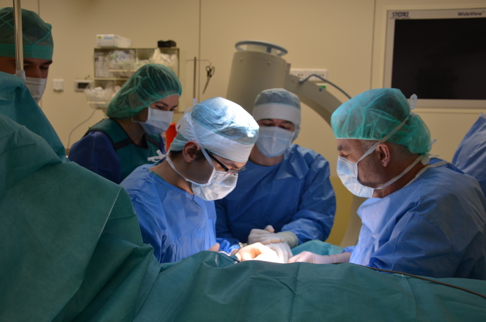
performed procedure, on the right prof. Tomasz Mazurek
Preparations for the operation lasted several weeks and took place at ChM. They started with printing deformed bones in polymer (based on CT scans). Later, a virtual model of correctly reproduced bones was created and also printed. On this basis, also by means of virtual modelling, the places and angles of intersection of the bones were selected in such a way that they were correctly set after their reassembly. The final part was designed surgical tools needed for the operation and the implants themselves. The prototypes were printed in 3D printing technology from polymers. After the tests were performed on the prepared models, the necessary equipment was printed in titanium in SLM technology: special gauges, wires to block them during bone cutting, and appropriately modelled plates and screws used to reattach the cut bones.
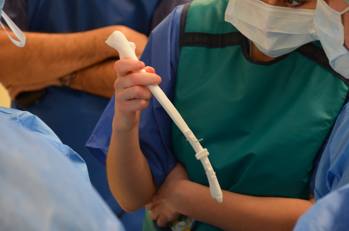
– The work carried out was interesting because, in parallel with the implant, special instruments were designed for this purpose. The whole philosophy of the surgical technique was based on an innovative tool, the so-called template with crosshair – explained Janusz Mroczkowski, ChM’s Spokesman.

tool printed for the procedure
Such operations are also possible by traditional methods. Based on the X-ray image, orthopaedists assess the type of deformation and determine the method of cutting the bone, and then during the operation they determine the appropriate bone angles and select the appropriate implant. However, these methods are imprecise and carry a significant risk of error. In addition, the lack of dedicated implants correcting a specific deformation requires the use of typical implants that are not always properly matched to the bone.
The use of modern technology allows the process to be planned before the patient arrives in the operating room. This shortens the duration of the procedure, substantially reduces the risk of complications and speeds up the recovery process after the procedure.
photo by Mikołaj Kopernik Hospital COPERNICUS in Gdańsk
Archives
- Academic Year 2024/2025
- Academic Year 2023/2024
- Academic Year 2022/2023
- Academic Year 2021/2022
- Academic Year 2020/2021
- Academic Year 2019/2020
- Academic Year 2018/2019
- Academic Year 2017/2018
- Academic Year 2016/2017
- Academic Year 2015/2016
- Academic Year 2014/2015
- Academic Year 2013/2014
- Academic Year 2012/2013